INTRODUCTION
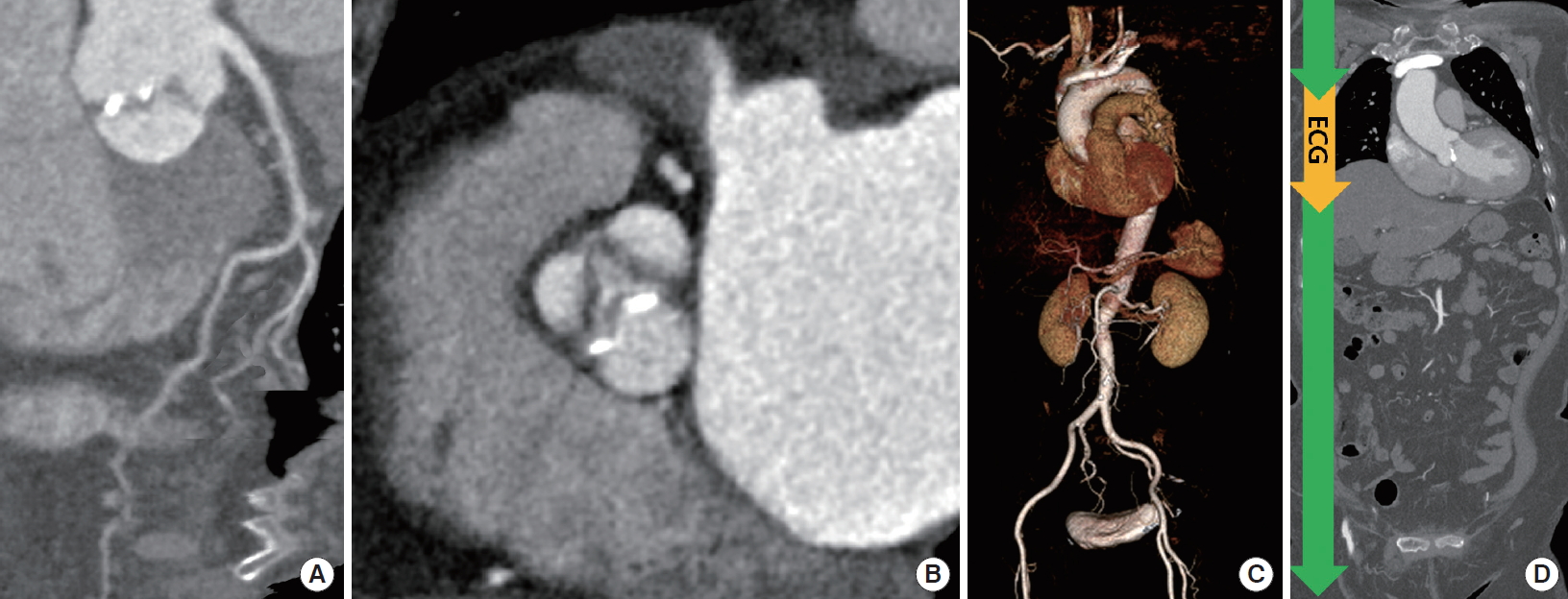
Recently, computed tomography (CT) and magnetic resonance imaging (MRI) are being utilized for the assessment of aortic valve disease (Figs. 1-7) [1-8]. Recent developments in CT enable simultaneous evaluation of the coronary arteries, aorta, proximal lower extremity arteries with the use of smaller contrast amount and lower radiation dose than before [9-17]. A wide-coverage (256- or 320-slice) detector design or high pitch dual-source CT enables a fast scan covering the aorta, coronary, and proximal femoral arteries (Fig. 1) [18]. Improved detector function and model-based iterative reconstruction are helpful in reducing the radiation dose of CT. Cardiac magnetic resonance (CMR) can be used to assess the severity of aortic stenosis (AS) and determine the prosthetic valve sizes as a reliable alternative to CT and transesophageal echocardiography (TEE) [6,19-21].
CORONARY ARTERY EVALUATION IN AS PATIENTS
Coronary computed tomography angiography (CCTA) may play a clinical role as an alternative to invasive coronary angiography (ICA) before cardiac valve surgery or transcatheter aortic valve implantation or replacement (TAVI/TAVR). With the high prevalence of coronary artery disease in patients with severe AS, percutaneous coronary intervention will become increasingly necessary in patients undergoing TAVI [22]. According to a meta-analysis of 13 studies, in 1,498 patients (mean age, 74years; 47% men; 76% TAVI procedures) undergoing surgical aortic valve surgery or TAVI, the pooled prevalence of significant stenosis (> 50%) determined by ICA was 43% [23]. CCTA showed the pooled sensitivity, specificity, and positive-likelihood and negative-likelihood ratios in detecting significant coronary stenosis, with ICA as the gold standard, of 95%, 79%, 4.48, and 0.06, respectively [23]. In a study involving 140 AS patients (68 males; 82.3±7.7 years) undergoing TAVI, the diagnostic performance of CCTA in the low calcium score group (42 [30%] patients,<400 Agatston calcium score) was better than that in the high calcium score group (98/140 [70%] patients, ≥400 calcium score) (area under receiver operating characteristic curve [AUC] 0.81 vs. 0.63) [24].
AS patients may have microvascular angina. Semi-quantitative myocardial perfusion reserve index (MPRI) using adenosine-stress CMR could detect impaired coronary microvascular function in patients with AS. According to a study of 117 patients with severe AS, MPRI values were much lower in the angina group than the asymptomatic group (0.74 ± 0.25 vs. 1.08 ± 0.28, P<0.001) and significantly lower in severe AS patients than in normal controls (0.90 ± 0.31 vs. 1.25 ± 0.21, P<0.001) [25]. As per logistic regression analysis, the only independent predictor for angina was MPRI (odds ratio, 0.003; P<0.001). On multivariate analysis, left ventricular mass index (LVMI) was found to be the strongest contributing factor to MPRI (standardization coefficient, –0.428; P<0.001) [25].
PRE- AND POST-TAVI EVALUATION WITH CT
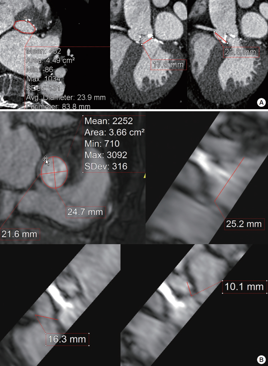
Recent CT techniques enable comprehensive evaluation before and after TAVI/TAVR in one breath hold with a variable or high helical pitch [7,9,13,14,16]. The CT area showed the highest correlation (0.932) among CT and TEE estimates, as compared with surgical annulus measurements [26].
The use of CCTA in addition to echocardiographic data in selecting the transcatheter heart valve (THV) size reduced the long-term occurrence of cardiovascular events [27]. Imaging strategy with CCTA was found to optimize and improve the outcome of TAVI. According to Casset et al. [27], multidetector-CT (MDCT) integration group improved the TAVI outcome compared with echocardiography only group (8% vs. 40%, P=0.008), mainly by decreasing the rate of paravalvular aortic regurgitation (PAR, 28% vs. 4%, P=0.04; major vascular complications 12% vs. 4%, P=0.6; all-cause death 16% vs. 4%, P=0.34; no stroke).
CCTA data and echocardiographic outcome data of the PARTNER II (Placement of AoRTic TraNscathetER Valves Trial II) SAPIEN 3 (Edwards Lifesciences, Irvine, CA, USA) intermediate-risk cohort of 835 patients showed that for the SAPIEN 3 THV, the frequency and extent of PAR was inversely related to the degree of oversizing, and acceptable rates of PAR were achieved at low degrees of oversizing [28]. Perimeter and area oversizing conferred similar predictive capacity in regard to the occurrence of PAR after THV implantation (AUC, 0.78 [95% CI, 0.70 to 0.85] vs. AUC, 0.78 [95% CI, 0.72 to 0.85]; P<0.001) [28]. In patients with area oversizing of ≥ 10%, moderate or high PAR was observed infrequently (0.3%). For low degrees of area oversizing, the rate of moderate or high PAR increased to 4.2% (for oversizing of ≥ 5% to 10%) and 2.8% (for oversizing of≥ 0% to 5%). The rate of moderate or high PAR increased to 7.8% for area undersizing of –5% to 0%, and further increased to 13.2% for undersizing below –5.0%. Similarly, a PAR rate of 2.4% was found for 0% to 2.5% perimeter oversizing and of 0.6% for≥ 2.5% perimeter oversizing. With perimeter undersizing down to –2.5%, the rate of moderate or high PAR increased to 6.8% and then further increased to 9.4% for pronounced undersizing of below –2.5%. The optimal cutoff for predicting moderate or high PAR was 0.05% for area oversizing and –1.24% for perimeter oversizing. Moderate or severe landing zone calcifications had a relative risk of 1.36 (95% CI, 1.09 to 1.69; P=0.006) for mild or higher PAR compared to none or mild landing zone calcifications.
Three-dimensional (3D)-printing based on CT data is a unique patient-specific method, allowing a tailored and individualized approach for aortic valve-sparing root reconstruction surgery [29]. With optimization, 3D models may help predict PAR in specific patients [30]. According to Ripley et al. [30], aortic root 3D models were highly accurate, with excellent agreement between annulus measurements made by 3D models and those made by corresponding 2D data (mean difference, −0.34 mm; 95% limits of agreement, ± 1.3 mm). Examination of the fit of valves within aortic root 3D models correctly predicted PAR in six of nine patients (six true positives, three false negatives) and absence of PAR in five of seven patients (five true negatives, two false positives).
MRI FOR TAVI EVALUATION
CMR is useful for TAVI evaluation before the procedure [31-33]. CMR can provide incremental information, including advanced tissue characterization with late gadolinium enhancement (LGE) and T1 mapping in patients with AS. In addition, left ventricle (LV) strain analysis with feature tracking and assessment of myocardial perfusion can be performed. CMR is now ready for implementation in clinical practice [20].
In the study of Cannao et al. [34], a non-contrast MRA protocol combining self-navigated 3D (SN3D) radial whole-heart and quiescent-interval single-shot (QISS) pulse sequences for the assessment of cardiac and vascular access route anatomy was technically feasible for TAVI planning. Acquisition time of the combined SN3D and QISS protocol was 10.1 ± 1.6 minute. All MRA measurements showed good agreement with CT angiography (CTA) in patients and there was no difference in qualitative ratings between MRA and CTA. Interobserver agreement was good for MRA (kappa =0.71 to 0.76) and excellent for CTA (kappa=0.82 to 0.84).
CMR is considered a reproducible, accurate, and reliable method to assess PAR severity as it allows direct quantification of AR. In a meta-analysis of seven studies by Papanastasiou et al. [35] on the level of agreement between two-dimensional transthoracic echocardiography (2D TTE) and CMR on grading the severity of aortic regurgitation (AR) after TAVI, six studies reported a low correlation between 2D TTE and CMR (kappa coefficient ranging from –0.02 to 0.41), whereas one study showed good agreement with a kappa coefficient of 0.72. The area under the curve for detection of moderate or severe AR with TTE was 0.83 (95% confidence interval [CI], 0.79 to 0.86), with CMR as the gold standard [35]. In the study by Salaun et al. [33], PAR severity was assessed 5 days after TAVI using TTE and CMR in 30 patients (COREVALVE, n = 10; EDWARDS SAPIEN XT, n= 20). In CMR, regurgitation volume and regurgitation fraction (RF) were significantly correlated with AR severity at TTE (P<0.001), with mean RF values of 9.2% ± 7.6% in mild (n = 22), 20.3% ± 4.2% in moderate (n =3), and 46.8% ±10.8% in severe PAR patients (n =5). A cutoff value of RF <14% on CMR accurately differentiated mild from moderate/severe PAR (sensitivity 100%, specificity 82%) [33].
AS EVALUATION WITH ECHOCARDIOGRAPHY, MRI, OR CT
Limitations of echocardiography
As per current guidelines, severe AS is defined as the aortic valve area normalized to body surface area (AVA/BSA) < 0.6 cm2/m2. According to Tribouilloy et al. [36], AVA/height showed better predictive performance than AVA/BSA with better reclassification and discrimination (net reclassification improvement, 0.33 vs. 0.28; integrated discrimination improvement, 0.10 vs. 0.08; C statistic, 0.67 vs. 0.65) than AVA/weight and AVA/body mass index. Inconsistency of hemodynamic characterization in severe AS between echocardiography (AVA < 1.0 cm2) and catheterization (mean pressure gradient > 40 mm Hg) is observed in about half of the patients, and this is only partly explained by low-flow (low LV ejection fraction [EF]) conditions [37]. This discordance can be also explained by underestimation of left ventricular outflow tract (LVOT) diameter or velocity-time integral. Doppler assessment may fail to accurately measure AVA in low-gradient AS patients and the coexistence of low-flow and a para doxically preserved LVEF (PLF-LG AS), is a real and frequently occurring entity (5% to 20%) [38]. Projected AVA at normal flow rate (250 mL/sec) calculated with stress echocardiography using low-dose dobutamine is superior to traditional severity criteria (AVA < 1.0 cm2 and mean gradient ≥ 40 mm Hg) in unmasking severe AS and predicting outcomes in patients with reduced LVEF. However, AVAproj is not reliable if the increase in transvalvular flow rate is below 15% and dobutamine-stress echocardiography may not be able to induce a significant increase in flow rate [38].
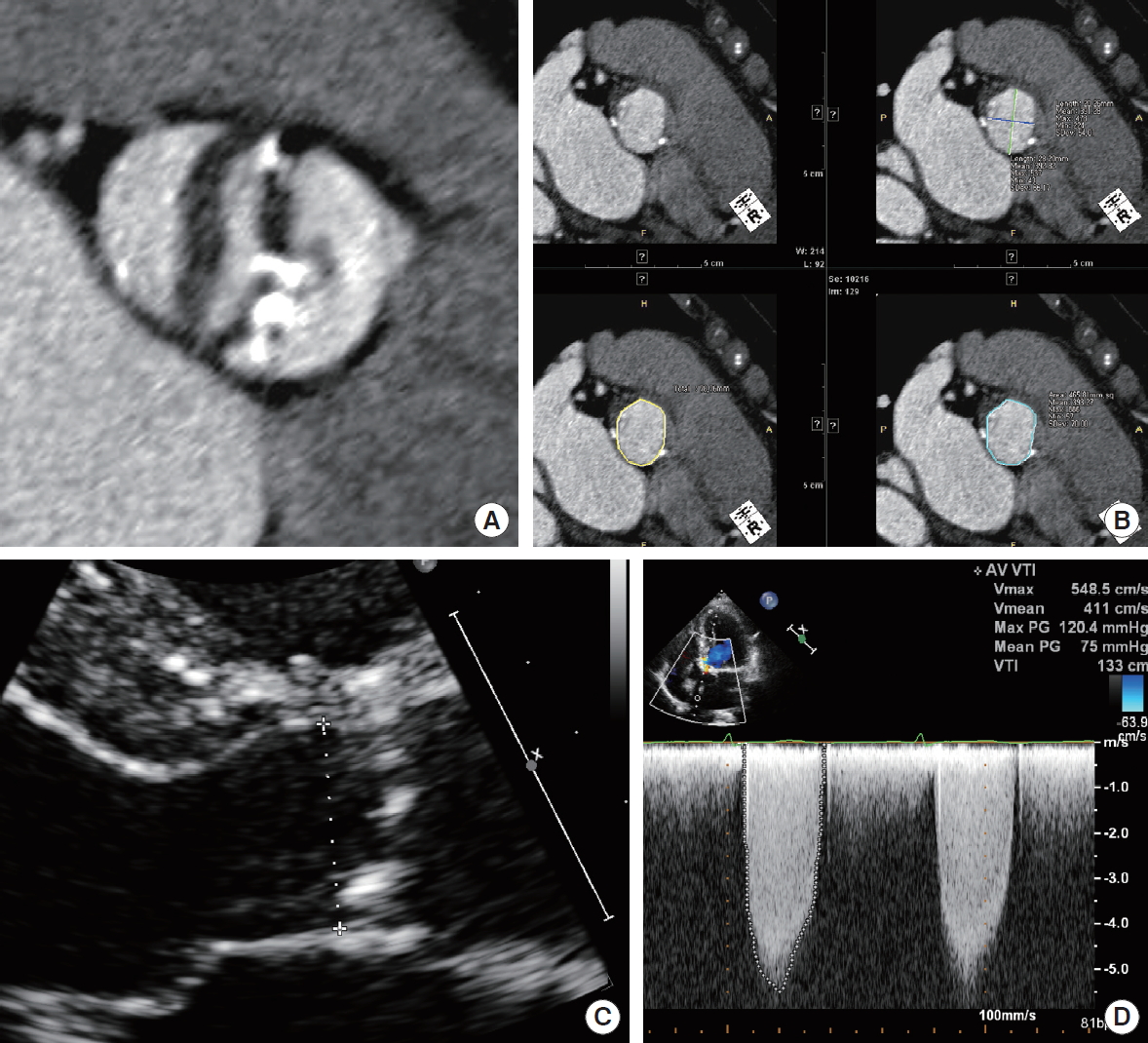
CT measurement of aortic valve area and calcium
According to the meta-analysis of nine studies (175 women and 262 men), MDCT is an efficient method for obtaining accurate AVA measurements in patients with AS [39]. The mean AVA as measured by CT was 1.0 ± 0.1 cm2 and the mean AVA measured by TTE was 0.9 ± 0.1 cm2 [39]. The correlation between CT and TTE AVA measurements was strong (r = 0.89) and the mean difference was 0.03 ± 0.05 cm2. Because of the elliptical anatomy of LVOT, 2D TTE may underestimate the LVOT area. Fusion of the LVOT area measured by CT and echocardiographic flow data has been proposed to improve the assessment of AS severity. For hemodynamic reasons, anatomic AVA is larger than effective AVA. Clavel et al. [40] calculated AVA corrected by LVOT area measured with CT using the following equation: AVACT = (LVOT area on CT)× VTILVOT/VTIAO, where VTILVOT is the velocity-time integral of LVOT and VTIAO is the VTI of stenotic aortic valve. In a study of 269 patients (76 ± 11 years of age, 61% men) with isolated calcific AS (mean gradient 44 ± 18 mm Hg; EF 58% ± 15%) who had undergone Doppler echocardiography and MDCT within the same episode of care, the head-to-head comparison of AVA with CT (AVACT) and with Doppler echocardiography (AVAEcho) revealed that AVACT was larger than AVAEcho (difference, 0.12 ± 0.16 cm2; P<0.001) but did not improve outcome prediction [40]. For long-term survival, after multivariable adjustment, AVAEcho and AVACT were independently predictive (hazard ratio [HR], 1.26; 95% CI, 1.13 to 1.42; P<0.001 vs. HR, 1.18; 95% CI, 1.09 to 1.29 per 0.10 cm2 decrease, respectively; P<0.001) with a similar prognostic value (P≥0.80) [40]. Large cut-point values should be used for severe AS, if AVACT (< 1.2 cm2) is compared with AVAEcho (< 1.0 cm2) [40].
Jander et al. [41] compared effective AVA (calculated from the continuity equation using CT-LVOT and TTE Doppler measurements) with anatomic AVA based on CT planimetry findings of 244 consecutive patients (mean age, 81 ± 5 years; 61% female) with AS. Substituting the LVOT area from TTE by the CT-LVOT resulted in an increase in AVA from 0.74 ± 0.15 to 0.92 ± 0.18 cm2 (P<0.01), which was larger than anatomic AVA from CT (0.82 ± 0.15 cm2) and AVA from TEE planimetry (0.79±0.14 cm2, P<0.01 vs. CT-LVOT). In the subgroup (n= 67) presenting with low-gradient severe AS and preserved EF, the results were similar (AVA from TTE, 0.76 ± 0.09; from CT-LVOT, 0.97 ± 0.14; CT planimetry, 0.86 ± 0.12; TEE planimetry, 0.82 ± 0.13 cm2). Aortic valve calcification (AVC) score quantitated by CT is helpful for identifying true severe AS by applying thresholds of 2,000 and 1,200 arbitrary units, respectively, for men and women. This modality should be considered, particularly if stress echocardiography is either not feasible or inconclusive [38]. Sex-specific CT-AVC thresholds enable identification of severe AS and provide prognostic information. In a multicenter study of 918 patients, CT-AVC provided excellent discrimination for severe AS (C statistic: women 0.92, men 0.89). The optimal sex-specific CT-AVC thresholds (Agatston score: women 1,377, men 2,062) were nearly identical to those previously reported (Agatston score: women 1,274, men 2,065) [42]. Sex-specific CT-AVC thresholds independently predicted aortic valve replacement (AVR) and death (HR, 3.90; 95% CI, 2.19 to 6.78; P<0.001) after adjustment for age, sex, peak velocity, and AVA in 215 patients with available clinical outcomes [42].
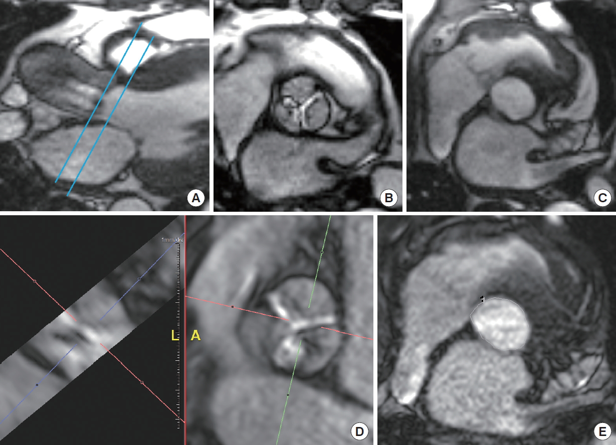
CMR assessment of AS severity
According to a systemic review, eight of 12 papers found CMR for the diagnosis of AS to have excellent reliability and reproducibility (low inter- and intraobserver variability) [2]. One study noted a sensitivity and specificity of 96% and 100%, respectively, when using CMR to detect AS diagnosed by cardiac catheterization (CC). Another study noted a lower sensitivity and specificity of 78% and 89%, respectively, which were better than the values reported when using TEE or TTE to detect severe AS, as noted on CC.
A head-to-head comparison between cine CMR and echocardiography for grading of AS severity revealed similar results between them, where AVA was the largest with planimetric CMR (0.93 ± 0.42 cm2) (TEE, 0.92 ± 0.32; continuity equation by CMR, 0.86 ± 0.30; TTE, 0.78 ± 0.25 cm2) [43]. CMR planimetry strongly correlated with TEE planimetry (concordance correlation coefficient [CCC], 0.85; 95% CI, 0.75 to 0.91). The aortic valve effective orifice area obtained by the continuity equation CMR was similar to that obtained by TTE (CCC, 0.82; 95% CI, 0.68 to 0.90).
According to Da Silveira et al. [44], multi-directional flow MRI, which encodes all three components of the velocity vector, can potentially outperform TTE in AS patients with eccentric or multiple jets. They compared fast 3-direction (3Dir) and 1-direction (1Dir) phase-contrast (PC) and TTE. The correlations ranged from 0.61 to 0.81 between TTE and 1Dir-PCCMR parameters and from 0.61 to 0.87 between TTE and 3Dir-PC CMR parameters. The correlation coefficients between TTE and 1Dir and 3Dir-PC CMR Vpeak were 0.81 and 0.87, respectively. Despite that, the comparison of Pearson’s correlations of the techniques did not reach statistical significance, even in the sub-analysis of severe cases (P> 0.05) [44].
Significant overestimation of transvalvular peak pressure drops occur with Doppler echocardiography because of approximation of blood flow as a single streamline. Donati et al. [45] assessed the accuracy of the Bernoulli principle in estimating the peak pressure drop at aortic valve using 3D CMR flow data in 32 subjects. Analysis of the pressure components confirmed that the spatial acceleration of the blood jet through the valve is the most significant aspect (accounting for 99% of the total drop in stenotic subjects). However, the Bernoulli formulation showed a consistent overestimation of the transvalvular pressure (average of 54%; range, 5% to 136%) resulting from the use of a single peak velocity value, which neglects the velocity distribution across the aortic valve plane. This assumption was the source of uncontrolled variability. A corrected formulation that accounts for the cross-sectional profile of the blood flow was proposed by the authors and adapted to both CMR and echocardiographic data [45].
MYOCARDIAL FIBROSIS
Myocardial fibrosis (MF) is regarded as an early and objective marker of left ventricular decompensation, particularly in asymptomatic AS patients [46]. Replacement MF or diffuse MF can be evaluated by LGE and T1 mapping [5,47-50]. Treibel et al. [51] investigated MF in 133 patients with severe, symptomatic AS using invasive biopsy and non-invasive imaging. Three patterns of MF were identified: (1) thickened endocardium with a fibrotic layer, (2) microscopic scars, with a subendomyocardial predominance, and (3) diffuse interstitial fibrosis. Collagen volume fraction (CVF) was elevated (P<0.001), as compared with controls, and higher (P<0.001) in endocardium-containing samples, with a decreasing CVF gradient from the subendocardium (P=0.001). LGE in grams, by the 3-standard deviation method, correlated with CVF (P<0.001) but not ECV. Both LGE and extracellular volume (ECV) correlated independently (P<0.001) with N-terminal pro-brain natriuretic peptide and high-sensitivity troponin T levels. Combined high ECV and LGE better identified patients with adverse LV remodeling, blood biomarkers, and histological parameters, and functional capacity than each parameter alone. A combined, multi-parametric approach with ECV and LGE allows efficient stratification of AS patients according to the response of the myocardial collagen matrix [51].
CMR techniques may be more sensitive than the conventional measures (LVEF or LV dimensions) to detect structural and functional changes in patients with severe left-sided valvular heart disease. The presence of MF has been associated with little improvement in clinical symptoms and recovery of LV systolic function [50]. On the contrary, Everett et al. [47] suggested that cellular hypertrophy and diffuse fibrosis (mid-wall LGE) progress in a rapid and balanced manner but are reversible after AVR in patients with AS. When mid-wall LGE is first identified, prompt AVR may improve clinical outcomes. According to a prospective trial by Everett et al. [47], in 61 asymptomatic patients (43% mild, 34% moderate, and 23% severe AS), significant increases in peak aortic jet velocity, LVMI, indexed ECV, and LGE mass were observed after 2.1 ± 0.7 years, with the most rapid progression observed in patients with severe stenosis. Patients with baseline mid-wall LGE (n= 16 [26%], LGE mass of 2.5 g [0.8 to 4.8 ]) showed particularly rapid increases in scar burden (78% [50% to 158%] increase in LGE mass per year). In 38 symptomatic patients (age, 66 ± 8 years; 76% men) who had undergone AVR, there was a 19% (11% to 25%) reduction in LVMI (P<0.0001) and an 11% (4% to 16%) reduction in indexed ECV (P=0.003) 0.9 ±0.3 years after surgery. On the contrary, mid-wall LGE (n=10 [26%]; mass of 3.3 g [2.6 to 8.0]) did not change after AVR (n=10; 3.5 g [2.1 to 8.0]; P=0.23), with no evidence of regression up to 2 years.
According to data from the British Society of Cardiovascular Magnetic Resonance Valve Consortium, in patients with severe AS, LGE on CMR was independently associated with mortality; its presence was associated with a 2-fold increase in late mortality [52]. Of the 674 patients with severe AS (75 ± 14 years, 63% male; AVA, 0.38 ± 0.14 cm2/m2; mean gradient 46 ± 18 mm Hg; LVEF, 61.0% ± 16.7%), scars were present in 51% (18% infarct-pattern; 33% non-infarct) [52]. Management involved surgical (SAVR, n = 399) or transcatheter procedures (TAVI, n= 275). Scar independently predicted allcause (26.4% vs. 12.9%, P<0.001) and cardiovascular mortality (15.0% vs. 4.8%, P<0.001), regardless of intervention (TAVI, P=0.002; SAVR, P=0.026 [all-cause mortality]). Every 1% increase in LV myocardial scar burden was associated with 11% increase in all-cause mortality (HR, 1.11; 95% CI, 1.05 to 1.17; P<0.001) and 8% increase in cardiovascular mortality (HR, 1.08; 95% CI, 1.01 to 1.17; P<0.001).
LV STRAIN
LV strain can be measured with CMR [49,53-56]. As a simple and practical method, tissue tracking is a promising method for assessing strain and predicting reverse remodeling in severe AS, especially in patients with suboptimal echocardiographic image quality. TAVI and SAVR procedures are associated with comparable decline in rotational LV mechanics at 6 months, with largely unchanged strain and strain rates [56]. On multivariable Cox analysis, baseline middle LV circumferential strain was significantly associated with all-cause mortality (HR, 1.03; 95% CI, 1.01 to 1.05; P=0.009), independent of age, LVEF, and Society of Thoracic Surgeons (STS) mortality risk score. Receiver operating curve analysis indicated that a mid-LV circumferential strain > –18.7% was associated with significantly reduced survival. No significant change in basal or middle LV circumferential strain or diastolic strain rate was seen after either intervention. However, a significant and comparable decline in LV torsion and twist was observed (SAVR: torsion, 14.08 ± 8.40 vs. 7.81 ± 4.51, P<0.001; twist, 16.17 ± 7.01 vs. 12.45 ± 4.78, P<0.01; TAVI: torsion, 14.43 ± 4.66 vs. 11.20 ± 4.62, P<0.001, twist, 16.08 ± 5.36 vs. 12.36 ± 5.21, P<0.001), which likely reflects an improvement towards normal physiology after alleviation of AS [56].
According to Hwang et al. [55], in 63 patients with severe AS and normal LV systolic function (EF > 60%), LV mass regression had significantly improved after AVR (baseline 145.9 ± 37.0 g/m2 vs. follow-up 97.7 ± 22.2 g/m2, P<0.001). Statistically significant Pearson’s correlations with LVMI regression were observed for longitudinal global strain (r = –0.461, P<0.001), radial strain (r= 0.391, P=0.002), and circumferential strain (r = –0.334, P=0.009). A simple linear regression analysis showed that all strain parameters could predict the amount of LVMI regression (P<0.05) as well as non-contrast T1 value (beta = –0.314, P<0.001) and ECV (beta = –2.546, P=0.038). However, ECV had the lowest predictive power (multiple r2= 0.071). Multiple regression analysis showed that strain could independently predict the amount of LVMI regression and the longitudinal global strain (beta = –3.335, P<0.001).
Using dedicated software, assessment of CT-derived LV strain is feasible. In patients treated with TAVI, CT-derived parameters of global myocardial strain can help improve short-term follow-up. Peak 3D global strain measured with CT in 25 TAVI patients showed increased peak global maximum principal strain after TAVI (0.59±0.18) from pre-TAVI values (0.46±0.19) [57].
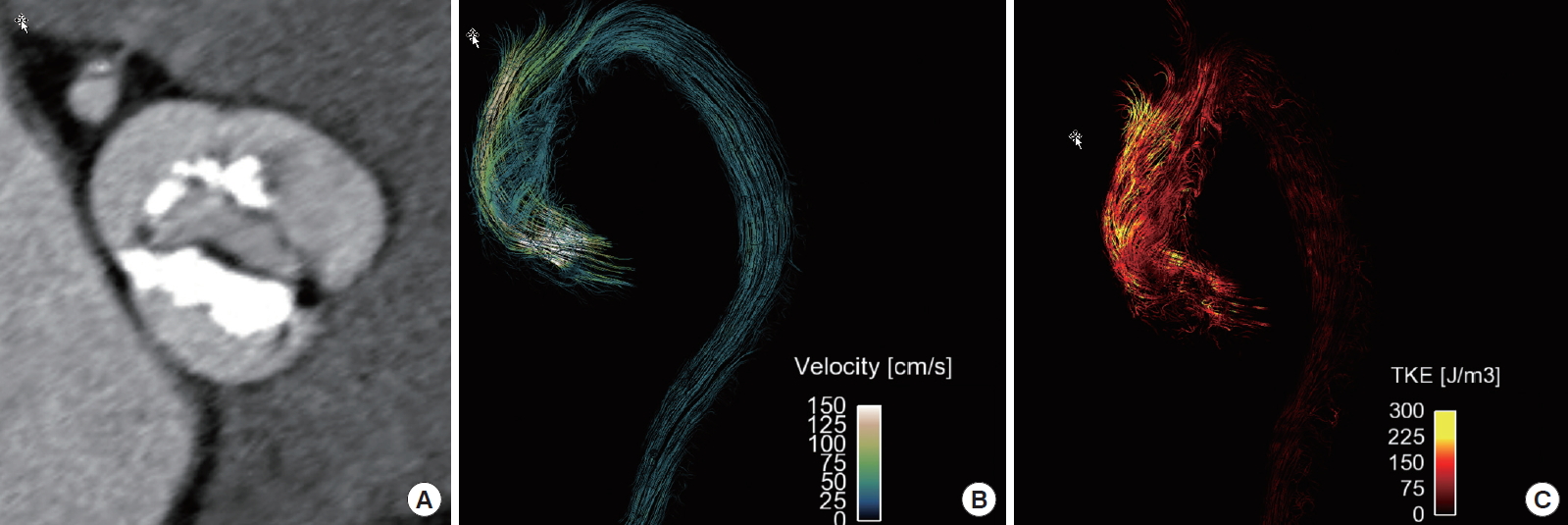
FOUR-DIMENSIONAL FLOW
Four-dimensional flow (4D flow) MRI applications for aortic valve disease provide an insight into aortic flow pattern, kinetic energy, wall shear stress (WSS) before and after TAVI, and aortic stiffness [3,45,58-64]. 4D flow MRI recorded significantly higher velocities than 2D phase-contrast MRI (2.04 ± 0.71 m/sec vs. 1.69 ± 0.79 m/sec, 17.2% difference, P<0.001) and similar velocities as Doppler echocardiography [65].
According to the study of Van Ooij et al. [66], AS significantly alters aortic hemodynamics and WSS independent of aortic valve phenotype. In all, 571 subjects underwent 4D flow MRI in the thoracic aorta (210 cusp fusions for right-left [RL] bicuspid aortic valve [BAV], 60 for right-noncoronary [RN] BAV, 245 tricuspid aortic valve patients with aortic dilatation, and 56 healthy controls). In BAV patients without AS, the different cusp fusion phenotypes resulted in distinct differences in eccentric WSS elevation: RL-BAV patients exhibited an increase in WSS of 9% to 34% (P<0.001) at the aortic root and along the entire outer curvature of the ascending aorta (AAo), whereas RN-BAV patients showed 30% WSS increase (P<0.001) at the distal portion of the AAo. WSS in tricuspid aortic valve patients with aortic dilatation without AS was significantly reduced by 21% to 33% (P<0.01) in four of six AAo regions. In all patient groups, mild, moderate, and severe AS resulted in a marked increase in regional WSS (P<0.001). Moderate-to-severe AS further increased WSS magnitude and variability in the AAo [66].
Rodriguez-Palomares et al. [61] analyzed differences in flow patterns and regional axial and circumferential WSS maps between BAV phenotypes and their correlation with AAo dilatation morphotypes. In all, 101 BAV patients (aortic diameter ≤45 mm, no severe valvular disease) and 20 healthy subjects were examined by 4D-flow CMR. BAV phenotype was RL in 78 patients and RN in 23. Both BAV phenotypes presented different outflow jet directions and velocity profiles that matched the location of maximum systolic axial WSS. RL-BAV velocity profiles and maximum axial WSS were homogeneously distributed right-anteriorly; however, RN-BAV showed variable profiles with a main proximal-posterior distribution shifting anteriorly at mid-distal AAo. Compared to controls, BAV patients presented similar WSS magnitude at proximal, middle, and distal AAo (P=0.764, P=0.516, and P=0.053, respectively) but low axial and high circumferential WSS components (P<0.001 for both, at all aortic levels). Different BAV phenotypes present different flow patterns with an anterior distribution in RL-BAV, whereas RN-BAV patients present a predominant posterior outflow jet at the sinotubular junction that shifts to anterior or right anterior in the middle and distal AAo. Thus, RL-BAV patients present a high axial WSS at the aortic root while RN-BAV patients present a high circumferential WSS in middle and distal AAo. These results may explain the different AAo dilatation morphotypes in the BAV population [61].
Turbulent kinetic energy (TKE), assessed by 4D flow MRI, is a measure of energy loss in disturbed flow as it occurs in AS. According to Binter et al. [59], elevated TKE levels imply high energy losses associated with BAV and dilated AAo geometries. In their study, 51 patients were divided into two groups (severe and mild/moderate AS) according to their echocardiographic mean pressure gradient. TKE values were integrated over the aortic arch to obtain the peak TKE. Integrating over systole yielded total TKEsys and by normalizing for stroke volume, normalized TKEsys was obtained. In patients with dilated AAo, both the peak TKE and total TKEsys were significantly elevated (P <0.01), whereas the mean pressure gradient was significantly low (P<0.05). Patients with BAV also showed significantly increased TKE metrics (P<0.01) although no significant difference was found for mean pressure gradient [59].
TAVI alters AAo blood flow and WSS patterns [60]. Compared to the controls, in TAVI patients, abnormally elevated WSS was noted in 30% ± 10% of the AAo wall. Increased WSS was seen along the posterior mid-AAo and the anterior distal AAo in all TAVI patients. TAVI results in eccentric and displaced flow in the middle and distal AAo, whereas blood flow displacement in SAVR patients occurs only in the distal AAo.
CONCLUSION
In conclusion, CT plays an important role in pre-TAVI evaluation and exclusion of coronary artery disease in AS patients. MRI can provide information on myocardial perfusion and MF in aortic valve patients. The characterization of blood flow patterns with 4D flow MRI may provide insights on the hemodynamics in aortic valve and the aorta in AS patients.