Warning: mkdir(): Permission denied in /home/virtual/lib/view_data.php on line 86 Warning: fopen(/home/virtual/pfmjournal/journal/upload/ip_log/ip_log_2024-04.txt): failed to open stream: No such file or directory in /home/virtual/lib/view_data.php on line 88 Warning: fwrite() expects parameter 1 to be resource, boolean given in /home/virtual/lib/view_data.php on line 89 Utility of renal artery ultrasound for detecting stenosis or obstruction: A narrative review
Precis Future Med Search

CLOSE


Precis Future Med > Volume 7(2); 2023 > Article
Park: Utility of renal artery ultrasound for detecting stenosis or obstruction: A narrative review

Abstract

Acute renal failure can result from many conditions, in which renal artery stenosis (RAS) or renal artery obstruction (RAO) can be corrected with appropriate interventional or surgical procedures. The renal artery can be directly depicted with currently available ultrasound (US) scanners. Therefore, we can determine if there is RAS or RAO in patients with poor renal function using renal artery US. However, there are few reports about the utility of renal artery US in excluding stenosis or obstruction. The purpose of this review is to show clinical applications and diagnostic pitfalls of renal artery US in detecting RAS or RAO.

INTRODUCTION

Computed tomography (CT) [1-4] or magnetic resonance imaging (MRI) [5-8] angiography has been accepted for assessing the renal artery and is most commonly performed prior to donor nephrectomy [9-12]. However, this imaging modality is difficult to conduct in patients with poor renal function because intravenous injection of contrast material is essential. Iodine or gadolinium can result in contrast-induced nephrotoxcity toxicity [13-16] or nephrogenic systemic fibrosis [17-20].
Ongoing development of ultrasound (US) scanner technology has improved image resolution so that deep-seated organs can be assessed. Prior to 2000, several investigations reported that US has difficulties in assessing the renal artery for stenosis or obstruction [21-23]. Accordingly, they relied on intra-renal Doppler US in diagnosing renal artery stenosis (RAS) or renal artery obstruction (RAO). Currently, the renal artery can be directly depicted with currently available US scanners [24-27]. RAS or RAO can be precisely identified with renal artery US. This imaging modality does not require CT or MRI contrast material, which should be excreted from the kidney in patients with poor renal function. Even though US contrast materials can be used to assess RAS or RAO, microbubbles can be excreted to the pulmonary circulation [28-31]. Therefore, renal artery US is a safe screening examination for those with suspected RAS or RAO. However, there are few reports about how renal artery US can be applied or its limitations. The purpose of this review is to show clinical applications and diagnostic pitfalls of renal artery US in detecting stenosis or obstruction.

CLINICAL APPLICATIONS

Follow-up of RAS

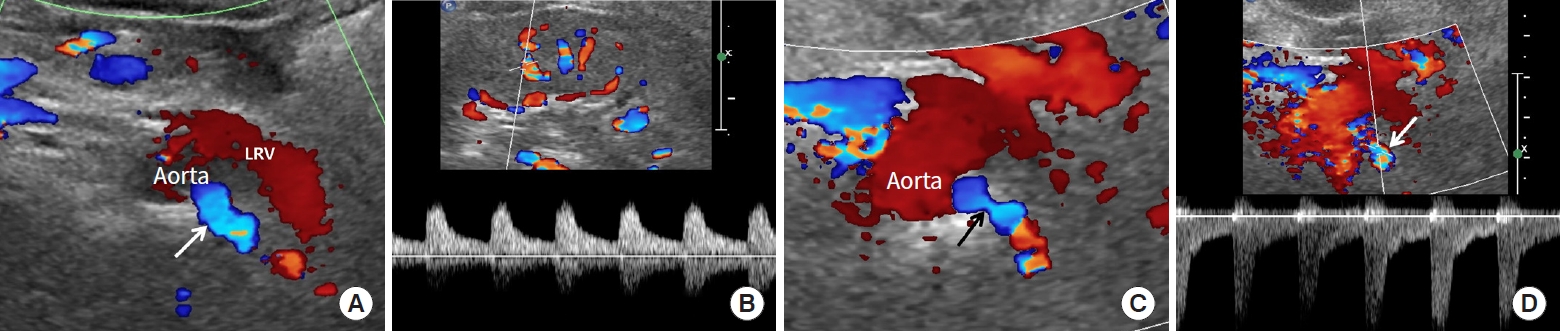
Renal artery US is useful for follow-up after angioplasty [32- 35] or stenting is performed [36-39]. It is important to identify re-stenosis when renal function or blood pressure is not well controlled during the follow-up period. The early stage of re-stenosis does not show pulsus tardus or parvus pattern in the kidney (Fig. 1). Therefore, the renal artery should be carefully evaluated to detect re-stenosis. Color or spectral Doppler US can provide key findings to determine re-stenosis following angioplasty or stenting (Fig. 1). Frequently, a renal artery stent obscures frequency shift from Doppler US due to posterior shadowing. If peak systolic velocity (PSV) is elevated in the renal artery distal to the stent, re-stenosis can be diagnosed. A large decrease in blood flow is another finding suggesting re-stenosis. Contrast-enhanced US can offer added value in depicting blood flow within a stent (Fig. 2).

Poor breath-holding

Breath-holding is necessary to obtain precise Doppler spectrum in the kidney. Accordingly, patient cooperation is essential during renal Doppler US. Many patients with RAS or RAO are old and/or have co-existing morbidities, and it is frequently difficult for them to hold their breath (Fig. 2). However, renal artery US does not require breath-holding for obtaining a renal artery Doppler spectrum (Fig. 2). First of all, the left renal vein should be searched with gray-scale US because renal arteries can be present behind it [24-27]. Then, gray-scale and Doppler US can depict RAS or RAO directly. As RAS becomes severe, the frequency shift is often not sufficient for detection. Thus, post-stenotic dilatation should be determined on a Doppler spectrum (Fig. 1). A PSV greater than 180 to 200 cm/sec in this area [24,40-44] or a PSV reno-aortic ratio (RAR) [44-47] greater than 3.5 strongly suggest RAS. The sensitivity and specificity of PSV > 180 to 200 cm/sec for RAS range from 85%–97% and 72%–98%, respectively [27,42,43,48]. The sensitivity and specificity of RAR > 3.5 for RAS range from 91%– 92% and 71%–95%, respectively [45-47].

Acute kidney injury

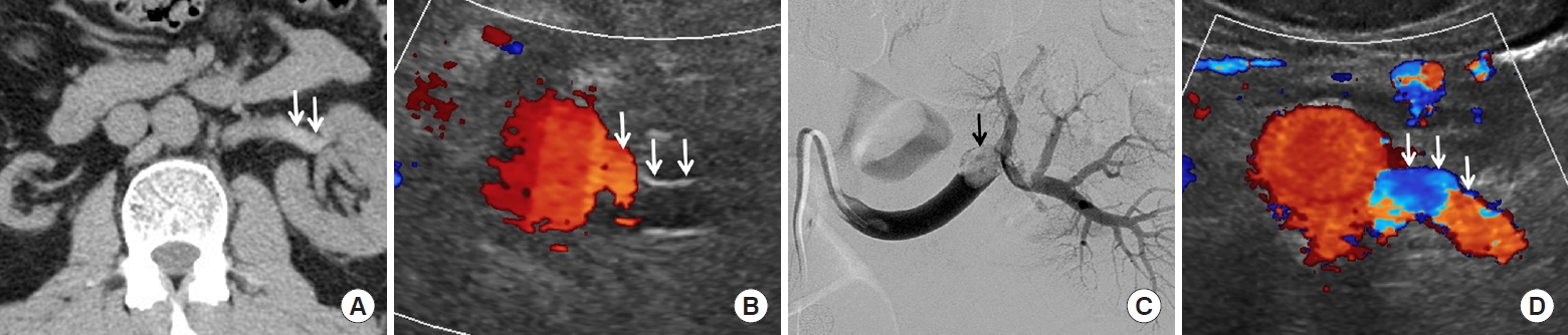
Contrast-enhanced CT or MRI is difficult to perform in patients with acute kidney injury (AKI) because of poor renal function [13-20]. Renal artery US helps to differentiate various conditions resulting AKI (Fig. 3). This US technique can exclude the possibility of RAS as an etiology of AKI. Therefore, if the renal artery is normal, radiologists should evaluate kidney size, echogenicity, perfusion, or obstruction with renal US to determine if AKI is pre-renal, renal, or post-renal [49-51]. For this reason, renal artery or renal US scan is recommended as a primary test for assessing patients with AKI rather than CT or MRI examination. A pre-renal etiology for AKI is most common [51]. The other etiologies include acute tubular necrosis, sepsis, hypovolemia, and drug toxicity [50,52]. Renal artery US helps nephrologists to manage AKI by excluding the likelihood of RAS [49-51].

Guidelines for angio-intervention

Renal artery US can offer good imaging features for detecting early stage RAS, for which angioplasty or stenting is a useful treatment. However, many investigations have reported that this angio-intervention is not as effective in treating chronic stage RAS, and that renal function or renovascular hypertension frequently is aggravated despite angio-intervention [53-56]. Subsequently, radiologists should be familiar with imaging features suggesting early stage RAS. As RAS becomes severe, arterial supply decreases and renal ischemia increases. As a result, cortical echogenicity increases and kidney size decreases. Intra-renal Doppler US shows decreased renal perfusion and pulsus tardus and parvus [57-59]. Early stage RAS can be defined when stenosis is present but kidney size and echogenicity are not significantly changed (Fig. 2). As a result, RAS should be detected before such anatomical changes are clearly depicted on US. When RAS is present in both renal arteries, renal artery US can indicate the treatment priority (Fig. 2). Radiologists should choose a renal artery based on kidney size or echogenicity.

Acute obstruction of renal artery

Acute obstruction of the renal artery results in acute renal infarction and rapid reduction of renal function [60-62]. CT or magnetic resonance angiography is limited for use in patients with poor renal function because of the need for intravenous contrast material. Renal artery US helps to determine RAO resulting from arterial dissection, acute thrombus, or surgical ligation (Fig. 3) [63,64]. These clinical conditions can lead to acute renal failure in patients with single kidney. RAO can occur if the renal artery is ligated during lymph node dissection around the aorta or intravenous vena cava. Early detection with renal artery US can minimize rapid loss of viable renal tissue resulting from acute RAO. Accordingly, this US technique can also help surgeons to determine treatment plans such as re-implantation of renal artery, auto-transplantation, or thrombolysis.

DIAGNOSTIC PITFALLS

Renal artery US has some limitations in assessing stenosis or obstruction. First, in patients with multiple renal arteries, special techniques are needed to detect or assess each renal artery [21-23]. If RAS occurs in the segmental renal artery, beginners might miss it on renal artery US. Even US experts should pay attention to carefully determine the number of renal arteries. If an aberrant renal artery is far from the origin of the main renal artery, it can be difficult to detect.
Second, a PSV greater than 180 to 200 cm/sec or an RAR greater than 3.5 on renal artery US strongly suggest RAS. However, patients with single kidney resulting from nephrectomy have a high PSV or RAR even though RAS is not present [65]. This is a physiologic phenomenon in which a single kidney needs to increase the glomerular filtration rate to match that of two kidneys. Therefore, PSV in a renal artery of a single kidney is almost twice that in paired kidneys immediately after unilateral nephrectomy. This physiologic finding should be differentiated with RAS.
Third, a similar finding can be achieved in the early period of renal allograft. PSV or RAR is higher in the early post-transplantation period than in native renal artery (Fig. 4) [66,67]. Blood flow is markedly elevated in the transplanted renal artery, and pseudo-RAS can be detected in renal artery anastomosis. Because the diameter of the recipient artery is larger than that of the donor artery, anastomosis frequently appears stenotic. Therefore, this anatomical deformity should be differentiated from RAS.
Fourth, renal artery US can detect RAS in the main or segmental renal artery. However, this US technique cannot easily detect small-vessel diseases in the subsegmental renal arteries due to limited image resolution [26]. This limitation is also found in CT or magnetic resonance angiography. Therefore, if small-vessel renal disease is suspected, conventional angiography is a management of choice.

SUMMARY

Renal artery US is useful in assessing poor renal function in patients who have poor breath-holding, follow-up after angio-intervention, screening for AKI, guidelines to angio-intervention, and acute obstruction of the renal artery. However, this US technique has limitations in patients who have multiple renal arteries, single kidney, allograft renal artery, or small-vessel disease.

CONCLUSION

Renal artery US can help to detect RAS or RAO in those in whom their renal function becomes poor. Therefore, radiologists or sonographers need to be familiar with this imaging techniques and imaging features.

CONFLICTS OF INTEREST

No potential conflict of interest relevant to this article was reported.

Notes

AUTHOR CONTRIBUTIONS

Conception or design: BKP.

Acquisition, analysis, or interpretation of data: BKP.

Drafting the work or revising: BKP.

Final approval of the manuscript: BKP.

Fig. 1.
Follow-up renal artery ultrasound (US) in a 66-year-old woman with angioplasty. (A) Color Doppler US shows no sign of re-stenosis in the left renal artery (white arrow) behind the left renal vein (LRV). She underwent angioplasty of the left renal artery stenosis 1 month prior. (B) Spectral Doppler US does not show pulsus tardus and parvus in the left kidney. This US scan was performed 2 years after the last angioplasty due to decreased renal function. There was no sign of re-stenosis on renal Doppler US. (C) Color Doppler US shows clear restenosis (black arrow) in the left renal artery. (D) Spectral Doppler US shows a high peak systolic velocity (270 cm/sec) in the post-stenotic area (white arrow). Systolic velocity is difficult to measure in the stenosis, in which the frequency shift is too small to detect. Subsequent angiography confirmed re-stenosis, and angioplasty was repeated.
pfm-2023-00009f1.jpg
Fig. 2.
Poor breath-holding in an 81-year-old woman with bilateral renal artery stenosis. (A) Color Doppler ultrasound (US) shows markedly decreased perfusion in the right kidney. Spectral Doppler US could not be obtained due to poor breath-holding in addition to poor renal perfusion. The size of the right kidney was 8.5 cm. (B) Gray-scale US shows smaller left kidney (7.5 cm), in which the left renal cortex is thinner and hyperechoic than those in the right. (C) Color Doppler US shows focal stenosis (white arrow) and post-stenotic dilatation (asterisk) in the right renal artery. (D) Color Doppler US shows another stenosis (white arrow) and post-stenotic dilatation (asterisk) in the left renal artery. The left renal artery stenosis is more severe, but post-stenotic dilatation is smaller compared to those in the right. The right renal artery is larger than the left. (E) Aortography shows clear bilateral renal artery stenoses (white arrows) and post-stenotic dilatations (asterisks). The stenotic degree is larger, but the post-stenotic dilatation is smaller in the right renal artery than in the left. The right renal artery is larger than the left. After considering imaging features of renal artery US and aortography, we decided to conduct angio-intervention in the right renal artery because it appears more effective in controlling renal function or blood pressure. (F) Angioplasty and stenting (white arrow) are successfully performed to dilate stenosis in the right renal artery alone. (G) Contrast-enhanced US shows the morphologic changes of right renal artery stenosis before and after angio-intervention (white arrow). This US technique clearly shows good blood flow within the stent.
pfm-2023-00009f2.jpg
Fig. 3.
Acute obstruction of the left renal artery in a 62-year-old man. (A) Non-contrast computed tomography (CT) shows a hyperdense sign (white arrows) in the distal left renal artery, suggesting acute thrombus. His renal function relies on a single left kidney because the right kidney is severely atrophic. (B) Color Doppler ultrasound (US) shows very weak blood flow in the proximal left renal artery (white arrows). (C) Arteriography shows acute thrombus (black arrow) as a filling defect in the left renal artery. Thrombolysis was performed to treat it. (D) Color Doppler US shows re-canalized left renal artery (white arrows), in which good blood flow is restored after thrombolysis.
pfm-2023-00009f3.jpg
Fig. 4.
High peak systolic velocity in a 67-year-old woman with renal allograft. (A) Spectral Doppler ultrasound (US) shows a high peak velocity (223 cm/sec) within the renal artery (arrow) of a renal allograft. This US scan was performed 3 days after the renal allograft was transplanted. (B) Right iliac arteriography shows no sign of stenosis in the allograft renal artery (arrow).
pfm-2023-00009f4.jpg

REFERENCES

1. Berg MH, Manninen HI, Vanninen RL, Vainio PA, Soimakallio S. Assessment of renal artery stenosis with CT angiography: usefulness of multiplanar reformation, quantitative stenosis measurements, and densitometric analysis of renal parenchymal enhancement as adjuncts to MIP film reading. J Comput Assist Tomogr 1998;22:533–40.
crossref pmid
2. Johnson PT, Halpern EJ, Kuszyk BS, Heath DG, Wechsler RJ, Nazarian LN, et al. Renal artery stenosis: CT angiography: comparison of real-time volume-rendering and maximum intensity projection algorithms. Radiology 1999;211:337–43.
crossref pmid
3. Urban BA, Ratner LE, Fishman EK. Three-dimensional volume-rendered CT angiography of the renal arteries and veins: normal anatomy, variants, and clinical applications. Radiographics 2001;21:373–86.
crossref pmid
4. Cho ES, Yu JS, Ahn JH, Kim JH, Chung JJ, Lee HK, et al. CT angiography of the renal arteries: comparison of lower-tube-voltage CTA with moderate-concentration iodinated contrast material and conventional CTA. AJR Am J Roentgenol 2012;199:96–102.
crossref pmid
5. King BF Jr. MR angiography of the renal arteries. Semin Ultrasound CT MR 1996;17:398–403.
crossref pmid
6. Dong Q, Schoenberg SO, Carlos RC, Neimatallah M, Cho KJ, Williams DM, et al. Diagnosis of renal vascular disease with MR angiography. Radiographics 1999;19:1535–54.
crossref pmid
7. Zhang H, Schoenberg S, Prince MR. MR angiography of the renal arteries. In: Schneider G, Prince MR, Meaney JFM, Ho VB, editors. Magnetic resonance angiography: techniques, indications and practical applications. Milano (IN): Springer Milan; 2005. p. 209–29.

8. Postma CT, Joosten FB, Rosenbusch G, Thien T. Magnetic resonance angiography has a high reliability in the detection of renal artery stenosis. Am J Hypertens 1997;10(9 Pt 1):957–63.
crossref pmid
9. Kim JK, Kim JH, Bae SJ, Cho KS. CT angiography for evaluation of living renal donors: comparison of four reconstruction methods. AJR Am J Roentgenol 2004;183:471–7.
crossref pmid
10. Chai JW, Lee W, Yin YH, Jae HJ, Chung JW, Kim HH, et al. CT angiography for living kidney donors: accuracy, cause of misinterpretation and prevalence of variation. Korean J Radiol 2008;9:333–9.
crossref pmid pmc
11. Chu LC, Sheth S, Segev DL, Montgomery RA, Fishman EK. Role of MDCT angiography in selection and presurgical planning of potential renal donors. AJR Am J Roentgenol 2012;199:1035–41.
crossref pmid
12. Arevalo Perez J, Gragera Torres F, Marin Toribio A, Koren Fernandez L, Hayoun C, Daimiel Naranjo I. Angio CT assessment of anatomical variants in renal vasculature: its importance in the living donor. Insights Imaging 2013;4:199–211.
crossref pmid pmc pdf
13. Mohammed NM, Mahfouz A, Achkar K, Rafie IM, Hajar R. Contrast-induced nephropathy. Heart Views 2013;14:106–16.
crossref pmid pmc
14. McCullough PA. Contrast-induced acute kidney injury. J Am Coll Cardiol 2008;51:1419–28.
crossref pmid
15. Solomon R, Dauerman HL. Contrast-induced acute kidney injury. Circulation 2010;122:2451–5.
crossref pmid
16. McCullough PA, Choi JP, Feghali GA, Schussler JM, Stoler RM, Vallabahn RC, et al. Contrast-induced acute kidney injury. J Am Coll Cardiol 2016;68:1465–73.
crossref pmid
17. Sadowski EA, Bennett LK, Chan MR, Wentland AL, Garrett AL, Garrett RW, et al. Nephrogenic systemic fibrosis: risk factors and incidence estimation. Radiology 2007;243:148–57.
crossref pmid
18. Kaewlai R, Abujudeh H. Nephrogenic systemic fibrosis. AJR Am J Roentgenol 2012;199:W17–23.
crossref pmid
19. Marckmann P, Skov L, Rossen K, Dupont A, Damholt MB, Heaf JG, et al. Nephrogenic systemic fibrosis: suspected causative role of gadodiamide used for contrast-enhanced magnetic resonance imaging. J Am Soc Nephrol 2006;17:2359–62.
pmid
20. Schlaudecker JD, Bernheisel CR. Gadolinium-associated nephrogenic systemic fibrosis. Am Fam Physician 2009;80:711–4.
pmid
21. Berland LL, Koslin DB, Routh WD, Keller FS. Renal artery stenosis: prospective evaluation of diagnosis with color duplex US compared with angiography. Work in progress. Radiology 1990;174:421–3.
crossref pmid
22. Desberg AL, Paushter DM, Lammert GK, Hale JC, Troy RB, Novick AC, et al. Renal artery stenosis: evaluation with color Doppler flow imaging. Radiology 1990;177:749–53.
crossref pmid
23. Hansen KJ, Tribble RW, Reavis SW, Canzanello VJ, Craven TE, Plonk GW Jr, et al. Renal duplex sonography: evaluation of clinical utility. J Vasc Surg 1990;12:227–36.
crossref
24. Granata A, Fiorini F, Andrulli S, Logias F, Gallieni M, Romano G, et al. Doppler ultrasound and renal artery stenosis: an overview. J Ultrasound 2009;12:133–43.
crossref pmid pmc
25. Schaberle W, Leyerer L, Schierling W, Pfister K. Ultrasound diagnostics of renal artery stenosis: stenosis criteria, CEUS and recurrent in-stent stenosis. Gefasschirurgie 2016;21:4–13.
pmid
26. Al-Katib S, Shetty M, Jafri SM, Jafri SZ. Radiologic assessment of native renal vasculature: a multimodality review. Radiographics 2017;37:136–56.
crossref pmid
27. Williams GJ, Macaskill P, Chan SF, Karplus TE, Yung W, Hodson EM, et al. Comparative accuracy of renal duplex sonographic parameters in the diagnosis of renal artery stenosis: paired and unpaired analysis. AJR Am J Roentgenol 2007;188:798–811.
crossref pmid
28. Ignee A, Atkinson NS, Schuessler G, Dietrich CF. Ultrasound contrast agents. Endosc Ultrasound 2016;5:355–62.
crossref pmid pmc
29. Park BK, Kim B, Kim SH, Ko K, Lee HM, Choi HY. Assessment of cystic renal masses based on Bosniak classification: comparison of CT and contrast-enhanced US. Eur J Radiol 2007;61:310–4.
crossref pmid
30. Melany ML, Grant EG, Duerinckx AJ, Watts TM, Levine BS. Ability of a phase shift US contrast agent to improve imaging of the main renal arteries. Radiology 1997;205:147–52.
crossref pmid
31. Drelich-Zbroja A, Jargiello T, Drelich G, LewandowskaStanek H, Szczerbo-Trojanowska M. Renal artery stenosis: value of contrast-enhanced ultrasonography. Abdom Imaging 2004;29:518–24.
crossref pmid pdf
32. Morellato C, Bergelin RO, Cantwell-Gab K, Zierler RE, Polissar NL, Davidson RC, et al. Clinical and duplex ultrasound follow-up after balloon angioplasty for atherosclerotic renal artery stenosis. Vasc Surg 2001;35:85–93.
crossref pmid pdf
33. Schaberle W, Strauss A, Neuerburg-Heusler D, Roth FJ. Value of duplex sonography in diagnosis of renal artery stenosis and its value in follow-up after angioplasty (PTA). Ultraschall Med 1992;13:271–6.
pmid
34. Taylor DC, Moneta GL, Strandness DE Jr. Follow-up of renal artery stenosis by duplex ultrasound. J Vasc Surg 1989;9:410–5.
crossref pmid
35. Tullis MJ, Zierler RE, Glickerman DJ, Bergelin RO, Cantwell-Gab K, Strandness DE Jr. Results of percutaneous transluminal angioplasty for atherosclerotic renal artery stenosis: a follow-up study with duplex ultrasonography. J Vasc Surg 1997;25:46–54.
crossref pmid
36. Chi YW, White CJ, Thornton S, Milani RV. Ultrasound velocity criteria for renal in-stent restenosis. J Vasc Surg 2009;50:119–23.
crossref pmid
37. Del Conde I, Galin ID, Trost B, Kang J, Lookstein R, Woodward M, et al. Renal artery duplex ultrasound criteria for the detection of significant in-stent restenosis. Catheter Cardiovasc Interv 2014;83:612–8.
crossref pmid
38. Mapes-Gonnella T. The role of sonography in renal artery stenosis evaluation and stent placement. J Diagn Med Sonogr 2013;29:78–86.
crossref pdf
39. Thornton SN, Cardenas GA, Imsais JK, Daniel GA, Afshar H, Goldman C, et al. Abstract 3308: New doppler ultrasound criteria for renal artery in-stent restenosis. Circulation 2007;116(Suppl 16):II-745.
crossref
40. Olin JW, Piedmonte MR, Young JR, DeAnna S, Grubb M, Childs MB. The utility of duplex ultrasound scanning of the renal arteries for diagnosing significant renal artery stenosis. Ann Intern Med 1995;122:833–8.
crossref pmid
41. Bokhari SW, Faxon DP. Current advances in the diagnosis and treatment of renal artery stenosis. Rev Cardiovasc Med 2004;5:204–15.
pmid
42. Radermacher J, Chavan A, Schaffer J, Stoess B, Vitzthum A, Kliem V, et al. Detection of significant renal artery stenosis with color Doppler sonography: combining extrarenal and intrarenal approaches to minimize technical failure. Clin Nephrol 2000;53:333–43.
pmid
43. Hua HT, Hood DB, Jensen CC, Hanks SE, Weaver FA. The use of colorflow duplex scanning to detect significant renal artery stenosis. Ann Vasc Surg 2000;14:118–24.
crossref pmid
44. Zierler RE, Bergelin RO, Davidson RC, Cantwell-Gab K, Polissar NL, Strandness DE Jr. A prospective study of disease progression in patients with atherosclerotic renal artery stenosis. Am J Hypertens 1996;9:1055–61.
crossref pmid
45. Aytac SK, Yigit H, Sancak T, Ozcan H. Correlation between the diameter of the main renal artery and the presence of an accessory renal artery: sonographic and angiographic evaluation. J Ultrasound Med 2003;22:433–9.
pmid
46. Bude RO, Forauer AR, Caoili EM, Nghiem HV. Is it necessary to study accessory arteries when screening the renal arteries for renovascular hypertension? Radiology 2003;226:411–6.
crossref pmid
47. Labropoulos N, Ayuste B, Leon LR Jr. Renovascular disease among patients referred for renal duplex ultrasonography. J Vasc Surg 2007;46:731–7.
crossref pmid
48. Hoffmann U, Edwards JM, Carter S, Goldman ML, Harley JD, Zaccardi MJ, et al. Role of duplex scanning for the detection of atherosclerotic renal artery disease. Kidney Int 1991;39:1232–9.
crossref pmid
49. Podoll A, Walther C, Finkel K. Clinical utility of gray scale renal ultrasound in acute kidney injury. BMC Nephrol 2013;14:188.
crossref pmid pmc pdf
50. Beaubien-Souligny W, Denault A, Robillard P, Desjardins G. The role of point-of-care ultrasound monitoring in cardiac surgical patients with acute kidney injury. J Cardiothorac Vasc Anesth 2019;33:2781–96.
crossref pmid
51. Kelahan LC, Desser TS, Troxell ML, Kamaya A. Ultrasound assessment of acute kidney injury. Ultrasound Q 2019;35:173–80.
crossref pmid
52. Hoste EA, Bagshaw SM, Bellomo R, Cely CM, Colman R, Cruz DN, et al. Epidemiology of acute kidney injury in critically ill patients: the multinational AKI-EPI study. Intensive Care Med 2015;41:1411–23.
crossref pmid pdf
53. Chrysochou C, Cheung CM, Durow M, Middleton RJ, Solomon LR, Craig A, et al. Proteinuria as a predictor of renal functional outcome after revascularization in atherosclerotic renovascular disease (ARVD). QJM 2009;102:283–8.
crossref pmid
54. White CJ. Optimizing outcomes for renal artery intervention. Circ Cardiovasc Interv 2010;3:184–92.
crossref pmid
55. Mukherjee D. Renal artery revascularization: is there a rationale to perform? JACC Cardiovasc Interv 2009;2:183–4.
pmid
56. Jennings CG, Houston JG, Severn A, Bell S, Mackenzie IS, Macdonald TM. Renal artery stenosis-when to screen, what to stent? Curr Atheroscler Rep 2014;16:416.
crossref pmid pmc pdf
57. Li JC, Yuan Y, Qin W, Wang L, Dai Q, Qi ZH, et al. Evaluation of the tardus-parvus pattern in patients with atherosclerotic and nonatherosclerotic renal artery stenosis. J Ultrasound Med 2007;26:419–26.
crossref pmid pdf
58. Ripolles T, Aliaga R, Morote V, Lonjedo E, Delgado F, Martinez MJ, et al. Utility of intrarenal Doppler ultrasound in the diagnosis of renal artery stenosis. Eur J Radiol 2001;40:54–63.
crossref pmid
59. Baxter GM, Aitchison F, Sheppard D, Moss JG, McLeod MJ, Harden PN, et al. Colour Doppler ultrasound in renal artery stenosis: intrarenal waveform analysis. Br J Radiol 1996;69:810–5.
crossref pmid
60. Silverberg D, Menes T, Rimon U, Salomon O, Halak M. Acute renal artery occlusion: presentation, treatment, and outcome. J Vasc Surg 2016;64:1026–32.
crossref pmid
61. Lessman RK, Johnson SF, Coburn JW, Kaufman JJ. Renal artery embolism: clinical features and long-term follow-up of 17 cases. Ann Intern Med 1978;89:477–82.
crossref pmid
62. Gasparini M, Hofmann R, Stoller M. Renal artery embolism: clinical features and therapeutic options. J Urol 1992;147:567–72.
crossref pmid
63. Cai S, Ouyang YS, Li JC, Dai Q, Tan L, Xia Y, et al. Evaluation of acute renal artery thrombosis or embolism with color Doppler sonography. Clin Imaging 2008;32:367–71.
crossref pmid
64. Walgraeve MS, Steyaert L, Gieraerts K. Contrast-enhanced ultrasound in a case of acute renal artery occlusion. J Belg Soc Radiol 2018;102:30.
crossref pmid pmc
65. Bohlouli A, Tarzamni MK, Zomorrodi A, Abdollahifard S, Hashemi B, Nezami N. Postnephrectomy changes in Doppler indexes of remnant kidney in unrelated kidney donors. Urol J 2009;6:194–8.
pmid
66. Patel U, Khaw KK, Hughes NC. Doppler ultrasound for detection of renal transplant artery stenosis-threshold peak systolic velocity needs to be higher in a low-risk or surveillance population. Clin Radiol 2003;58:772–7.
crossref pmid
67. Baxter GM, Ireland H, Moss JG, Harden PN, Junor BJ, Rodger RS, et al. Colour Doppler ultrasound in renal transplant artery stenosis: which Doppler index? Clin Radiol 1995;50:618–22.
crossref pmid


ABOUT
ARTICLES

Browse all articles >

ISSUES
TOPICS

Browse all articles >

EDITORIAL
POLICY
AUTHOR
INFORMATION
Editorial Office
Sungkyunkwan University School of Medicine
2066 Seobu-ro, Jangan-gu, Suwon, Gyeonggi-do 16419, Korea
Tel: +82-31-299-6038    Fax: +82-31-299-6029    E-mail: pfmjournal@skku.edu                

Copyright © 2024 by Sungkyunkwan University School of Medicine.

Developed in M2PI

Close layer
prev next